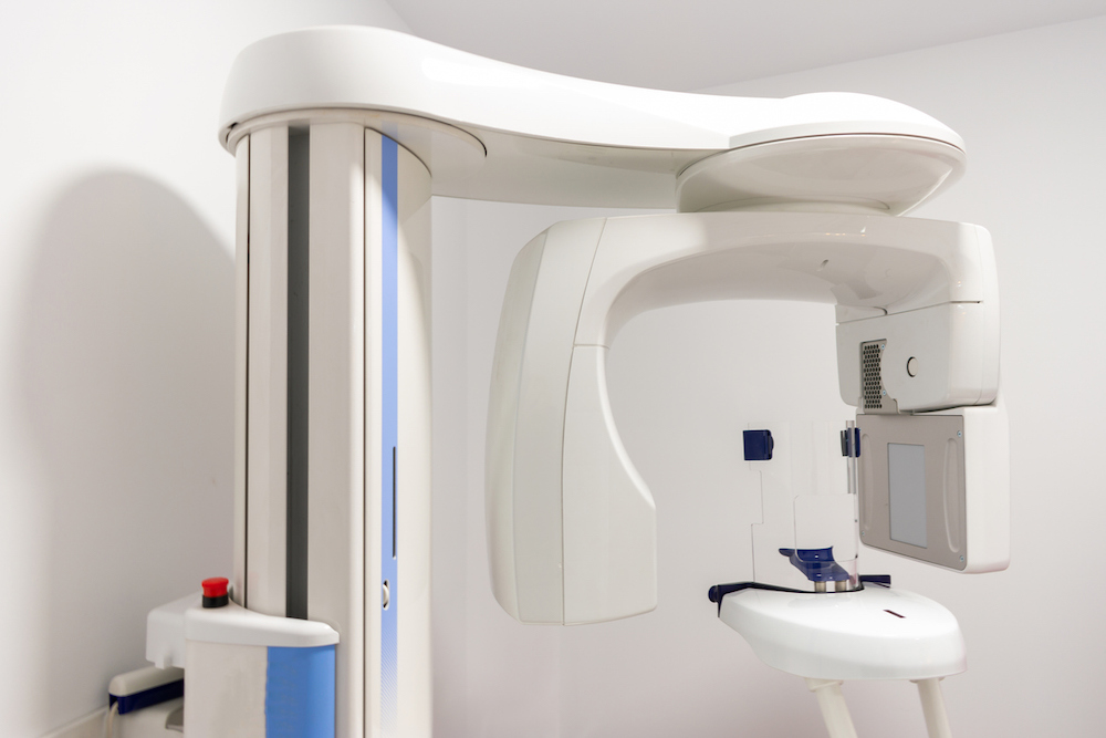
Using CBCT to measure dentin thickness of C-shaped root canal walls
“Danger zones” for C-shaped mandibular first and second premolars — where there is an increased risk of stripping perforation during endodontic preparation — are predominantly the mesial and distal walls of the lingual canal.
A retrospective, cross-sectional study published in Restorative Dentistry & Endodontics measured the dentin thickness of C-shaped canals in mandibular first and second premolars at coronal, middle and apical root levels using cone-beam computed tomography. Researchers found that the thickest walls for both premolars were the buccal and lingual walls at all three root levels. The thinnest walls were the mesial and distal walls of the lingual canal for the first premolars and the mesial end of the buccal and lingual canals for the second premolars.
C-shaped canal morphology is an anatomical variation in which independent root canals are merged or connected through isthmuses or fins, forming a C-shaped appearance at cross-sectional views. It is often associated with the fusion of the roots and a longitudinal radicular groove on the root surface.
The radicular groove is described as a developmental depression area that occurs because of root fusion and creates a danger zone. It has previously been reported that the root canal wall facing the radicular groove was thinner than the other walls in C-shaped mandibular molars, but fewer studies have described the presence of C-shaped canal morphology in mandibular premolars.
Read more: Restorative Dentistry & Endodontics
The article presented here is intended to inform you about the broader media perspective on dentistry, regardless of its alignment with the ADA's stance. It is important to note that publication of an article does not imply the ADA's endorsement, agreement, or promotion of its content.